Cross-section of paperboard with coating layer on top. SEM image by Torben Nilsson Pingel.
3D Imaging of Soft Materials
Soft materials are complex with hierarchical, multiscale, and multiphase structures. Since mass transport in these materials occurs in three dimensions, detailed 3D imaging is essential for accurate analysis and simulation.
Multiscale Imaging Techniques
To capture structures from nanometers to centimeters and with different contrast modes, a range of advanced microscopy methods can be combined, including optical microscopy, X-ray imaging and electron microscopy.
High-resolution in situ 3D imaging under varying conditions (e.g., temperature, humidity, applied force, electric fields) reveals dynamic structural changes critical to understanding transport processes.
TEM: Titan 80-300 at Chalmers University of Technology.
Quantitative Image Analysis
The 3D image data is analyzed to extract quantitative insights, linking structure to raw materials, manufacturing processes, and external influences.
By feeding the acquired 3D images into our in-house GPU-powered lattice Boltzmann software, simulations of the mass transport through these complex structures can be performed, with the results validated against experimental data.
Imaging Capabilities
Focused Ion Beam Scanning Electron Microscopy (FIB-SEM)
X-ray Tomography: Lab-scale and synchrotron facilities available
Transmission Electron Microscopy Tomography (TEMT): Ultra-high resolution 3D imaging
Confocal Laser Scanning Microscopy (CLSM): Micrometer-scale imaging
In addition, advanced sample preparation and cryo-techniques for microscopy are available.

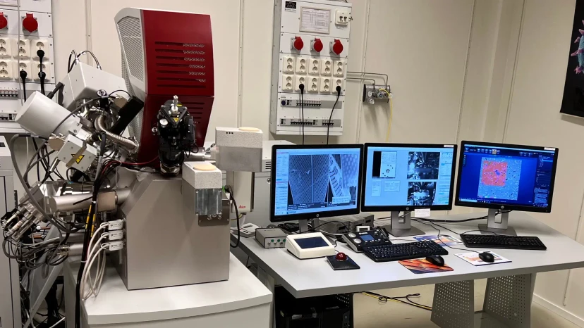
TEM: JEOL JEM-2100Plus at RISE Research Institutes of Sweden.
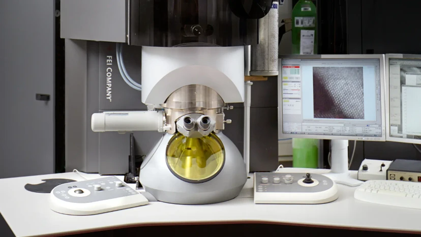
FIB-SEM: FEI Versa3D at Chalmers University of Technology.
Want to know more?
Please reach out to one of our experts in the field
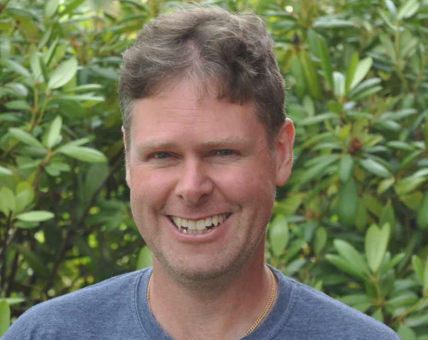
Senior researcher
Niklas Lorén
Senior scientist and Associate professor at RISE Research Institutes of Sweden. Expert in microstructure-property relationships in soft materials.
niklas.loren@ri.se+46 10 516 66 14

Professor
Eva Olsson
Professor at Deparment of Physics, Chalmers Universoty of Technology. Head of Division Nano and Biophysics.
eva.olsson@chalmers.se+46 31 772 32 47
Related publications
Explore some publications related to the concept.